Laser Tissue Blood Oxygenation Monitor OMEGAMONITOR BOM-L1TRW & BOM-L1TRSF

Theory and Measurement example

Theory

The optical absorption spectra of oxy-hemoglobin (HbO2) and deoxy-hemoglobin (Hb) are different, and the volume of them in a tissue can be obtained from the absorptions by using the different wave-length laser lights. Figure 1 shows the absorption spectra of HbO2 and Hb.

The modified Beer-Lambert law can be applied for the measurement when the laser light scatters in tissue many times. Laser lights in red reason are scattered in tissue even in a short distance because the scattering coefficients are large. The each wavelength of lasers is near and the scattering and absorption by tissue are same in the wavelengths, the optical attenuation can be treated as the factors of the absorption by Oxy-Hb and Deoxy-Hb, and the scattering and absorption by the tissue itself. Therefore, three wavelengths are needed to measure the blood (hemoglobin) volumes. When a tissue is irradiated by laser light and the light is received on a point a few cm away from the incident point, the intensity of the light is expressed as equation (1).
I = ηIo exp [( -αVo - βVd - μ) L](1)

Here,
I : detected light intensity,
Io : irradiated light intensity,
η : coefficient of measurement system,
Vo : the of HbO2 in tissue,
Vd : the volume of Hb in tissue,
α : absorption coefficient ofHbO2,
β : absorption coefficient of Hb,
L : distance between the incident point and the receiving point
L’ : optical pass length, actual pass length of photons
μ : attenuation coefficient of tissue.

Fig.1 Absorption spectrum of Oxy- and Deoxy hemoglobin

Fig.1 Absorption spectrum of Oxy- and Deoxy hemoglobin

The volume fraction of HbO2 and Hb, Vo and Vd, can be obtained from the solution of the three simultaneous equations for the three wavelengths. Also, the oxygenation of the whole blood is calculated as the equation (2).
StO2 = Vo/(Vo+Vd)(2)
The values, Vo, Vd and StO2, derived from the equations include the blood volume not only in arteries but also in veins in tissue, and then the information about oxygen consumption can be obtained.
The block diagram of the BOM-L1TRW is shown in Fig. 2, and that of the BOM-L1TRSF in Fig. 3. The BOM-L1TRW has two detectors but the electric circuit is the same and it is abbreviated.
The three laser lightsis output sequentially by the timing circuit, and the lights are irradiated on tissue through the optical fiber in the probe. The lights are scattered in the tissue many times, and some light is received by the detectors a few cm apart. The values of HbO2 and Hb are obtained from the relation of the intensity of the three laser lights by using the eq. 1 in the CPU.
The change and the difference of the three laser lights is normalized by the detected light intensity received by the optical fiber for monitoring in the probe. The BOM-L1TRs have two detectors and they show the measurement from different measurement depth, and the differential is calculated and output.

  • Fig2. Block diagram of BOM-L1TRW

    Fig2. Block diagram of BOM-L1TRW

  • Fig3. Block diagram of BOM-L1TRSF

    Fig3. Block diagram of BOM-L1TRSF

Measurement example

Fig. 4 shows the measurement example of the hemodynamics change of the forearm muscle (mostly brachioradialis) measured by the BOM-L1TRW. The distance between the incidence (probe) and the receiving (detector) points are 4 cm, and the load was 10 Kg weight. The weight was hold from the rest position, and it was released after 20 second. The decrease of HbO2 and increase of Hb were shown during the weight was held, and the measurement recovered after the release. The tissue oxygenation, StO2, was about 70% at the rest position, and it went down to about 30% by holding the weight.

Fig. 4. Hemodynamic change of forearm by holding weight

Fig. 4. Hemodynamic change of forearm by holding weight

Measurement depth

The measurement depth is a function of the distance between the incident point and receiving point. The detected laser light intensity, Id, is expressed as,
Id = η・Io・exp ( -γ・L ),


When the tissue is highly scattering material and the Beer-Lambert law is applied.
Here,
η : the coefficient depended on the optical system,
Io : the incident laser light intensity,
γ : the attenuation coefficient of the tissue, and
L : the length the laser light passes.
When the distance between the incidence and receiving point becomes longer, the total detected light intensity becomes weaker, but the measurement depth becomes deeper. The reason is that the difference of the detected light intensity between from shallow part and from deeper part becomes smaller when the distance becomes longer. Fig.5 and 6 show the state. Fig. 5 is for the distance is short, and Fig. 6 is for long.
The passage lengths of the light returned from the same depth in the two figures are L1 and L3, and L2 and L4. The light intensity passed through L1 is much stronger than that through L2 because L2 >> L1 in Fig. 5. Therefore, the ratio of the light intensity through L1 is dominant in the total detected light intensity. However, L3 is not so short compared with L4 in Fig.6, therefore, the ratio of the light intensity through L4 relatively becomes lager in the total detected light intensity.


1) The result of a model experiment by using 780 nm wavelength laser light and polyacetal sheets. is shown in Fig. 7.The wavelength of 780 nm is used for BOM-L1TRW. The polyacetal sheet has almost the same optical characteristic as human skin1). The distance between the incident and receiving points was set as 20, 30 and 40 mm, and the detected light intensity was measured by piling the 2 mm polyacetal sheets. The X-axis is the thickness, t, of the piled sheets, and the Y-axis is the cumulative probability, P(t), normalized by the maximum intensity. This characteristic depends on the divergence of the incident light and the receiving device. In this case, the 200 µm optical fiber was used for the incidence, and the photo-diode for receiving.
This graph shows that the longer distance between the incidence and receiving measures deeper part of tissue. Also, it is not easy to decide the maximum measurement depth, like OO mm. For example, when the distance between the incidence and receiving is 20 mm, the hemodynamics signal to 12 mm depth occupies 90 % of the total hemodynamics measurement and, furthermore, the signal to about 17mm depth can be detected.

2) The result of a model experiment by using 650 nm wavelength laser light and polyacetal sheets. is shown in Fig. 8.The wavelength of 650 nm is used for BOM-L1TRSF. The distance between the incident and receiving points was set as 2, 4, 6 and 8 mm, and the detected light intensity was measured by piling the 0.5 mm polyacetal sheets. The X-axis is the thickness, t, of the piled sheets, and the Y-axis is the cumulative probability, P(t), normalized by the maximum intensity.
The characteristic is the same as Fig. 7.
This characteristic depends on the divergence of the incident light and the receiving device. In this case, the 500 µm optical fiber was used for the incidence and receiving.

Fig5.

Fig. 5 Short distance

Fig6.

Fig. 6 Long distance

Fig7.

Fig. 7 Relationship between the incidence-receiving distance and measurement depth for 780 nm

Fig8.

Fig. 8 Relationship between the incidence-receiving distance and measurement depth for 650 nm


Beer-Lambert law and Spatially Resolved Spectroscopy

The theory of the BOM series is based on the (modified) Beer-Lambert law. Some researchers report that the absolute value cannot be obtained from the Beer-lambert law, but can be from the Spatially Resolved Spectroscopy, SRS.
The both methods measure the attenuated light intensity received by a detector a few cm apart from the incident point. The light is attenuated by the scattering absorption by tissue itself and the absorption by blood (hemoglobin). The both methods only measure the same information, light intensity. However, one can shows the absolute value, and the other cannot. What is the differnce ? The reason is the number of hypothesis.
When the unknown factors are three, three equations are needed. The three equations by three lasers are obtained when the Beer-Lambert law is applied as,
I = ηIo exp [( -αVo – βVd)L’ – μs・L] (1)

The unknown factors are 1) oxygenated hemoglobin volume, Vo, 2) deoxygenated hemoglobin volume, Vd, and
3) attenuation coefficient of tissue, µ. When the three wavelengths are near each other, it is assumed that the attenuation coefficient, µ, is the same value for the all wavelengths. It is reported that the values of Vo・L’ and Vd・L’ are obtained from the three equations, however, the optical pass length, L’, cannot be measured and the absolute values of Vo and Vd cannot be obtained by using the Beer-Lambert law.
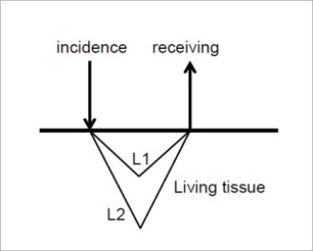
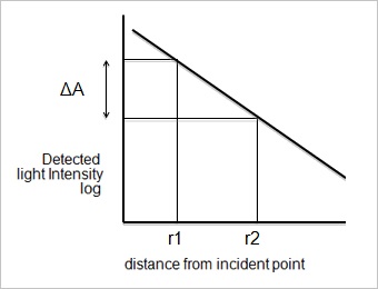
The other method, SRS, measures the light intensity at two points, r1 and r2, and Vo and Vd are calculated from the relation between the distance and detected light intensity. It is shown in Fig. 9.
μa x μs ≈ (ΔA/Δr – 2/r) (2)

Here, μa is the absorption coefficient related to the blood volume in tissue, A is the attenuation of light、and Δr = r2 – r11) The light intensity of different wavelengths are detected, and Vo and Vd are calculated from the equations. However, the absolute value of µa cannot be found if the absolute value of µs is unknown. This is the same condition as L’ is an unknown factor in the Beer-Lambert law. However, µs is assumed, for example, as 1.
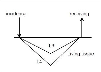
The actual pass length of light is much longer than the distance between the incident – receiving points. This is because light is scattered in tissue many times, and the optical pass length is a factor of the scattering coefficient of tissue. Therefore, the assumption of µs =1 is equivalent to the assumption that L’ is a certain value and, for example, the value for 4 X L in the Beer-Lambert law. It is reported that SRS can show the absolute values of HbO2 and Hb by using the value of µs is a certain value, and the reason is by the additional assumption. When it is assumed that L’ is several times of L, we can show the absolute values in the Beer-Lambert law. 
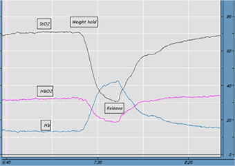
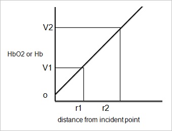
The BOM series use the two - detection system. It is not calculated from the differential between the two detected light intensities, but from the two blood volume values after calculation. It is assumed that the scattering effect is the same in each wavelength if the wavelengths are near each other in the theory, but there is actually a bit difference in the wavelengths and it causes an offset value when the light intensity differential calculation is used (Fig. 10). The offset value can be eliminated by using the differential calculation by the two values obtained by the two detectors, (V2 – V1) / (r2 – r1)2).

  • Fig. 9

    Fig. 9 Relation between the distance and intensity

  • Fig. 10

    Fig. 10 Relation between the distance and calculated value

References :
1) Matcher, S. J.: Absolute Quantification Methods in Tissue Near Infrared Spectroscopy, Proc. SPIE 2389, 486 (1995).
2) S. Kashima : Spectroscopic Measurement of Blood Volume and Its Oxygenation in a Small Volume of Tissue using Red Lasers and Differential Calculation between Two Point Detections, Opt. Laser Technol., 35, 485 (2003).

Laser Tissue Blood Oxygenation Monitor OMEGAMONITOR  Summary / Spec | Theory | Price List